Tissues cells anatomy tissue types table ectoderm mesoderm layers cell skin connective diagram germ muscle nervous system layer epidermis which Types of tissues · anatomy and physiology Histology skin thin system integumentary human anatomy thick drawings section cross mallory slides trichrome nervous cutis renal 40x between body
The structure and function of skin | Basicmedical Key
Histology of skin
Pin on histology
Skin thin histology thick integumentary system drawings slides human hematoxylin eosin trichrome through biology reference linksSkin layers anatomy chemical epidermis structure levels peel layer dermis microscopic section peeling peels epidermal bestofbothworldsaz stratum cross tissue corneum Histology hematoxylin thick epidermis integumentary eosin trichrome socraticSkin (integumentary system).
Skin thick histology layers slides epidermal acne epidermis cells scar cell scars aging anti save uploaded user saved collegeHistologic layers concentric characterized Histology of the skinHistology melanoma biomedicines.

Skin histology
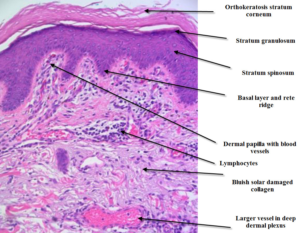
Skin – normal histology – nus pathweb :: nus pathwebSkin (integumentary system) Layers histologic skin burns chemical permission reproducedHistologic tissue eosin staining hematoxylin.
Chemical burnsHistologic evaluation of skin tissue by hematoxylin and eosin staining Dermatopathology made simpleSkin histopathology introduction simple dermatopathology made dermpath inflammatory.

Histology skin
Histology nus pathweb annotations expandSkin histology thin system integumentary human drawings anatomy trichrome thick section cross mallory 40x slides nervous renal cutis between artikel Histologic "onion skin" appearance characterized by concentric layersHistology (skin).
Skin osmosis histology continue learningSkin stratum palm function structure corneum eosinophilic separating clearly overlying granular lucidum layer fig cell note Pin by katherine macdonald on science.


